
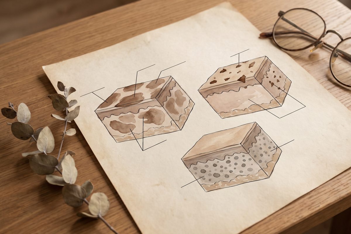
How does a sun spot differ from melasma?
A sun spot, properly a solar lentigo, is a benign flat mark caused by chronic sun exposure. It has a clear edge, a fairly even colour within the spot, and it turns up where the sun has reached for years: cheeks, temples, backs of the hands, the upper chest. Each spot is its own island. Most are addressed in a small number of visits, as the article on age spots explains.
Melasma looks like a shadow across the skin, with no separate islands. DermNet NZ describes bilateral light-to-dark brown patches with irregular borders, most often in a centrofacial pattern across the forehead, cheeks, nose and upper lip, in roughly half to four fifths of cases. It fluctuates. Sun, heat and hormonal shifts push it darker, which is why it is managed as a long-term condition and behaves less like a stain, and why the Malaysian climate keeps it active.
What is Hori's naevus, and why is it mistaken for melasma?
Hori's naevus is a dermal pigment condition. The pigment cells sit deeper than those of a sun spot, and light scattering through that depth gives the spots a grey or blue tinge on top of the brown. In a Vietnamese case series every patient had lesions on both cheeks and blue-brown was the commonest colour. A systematic review in 2026 describes it as a common dermal melanocytosis that mainly affects East Asian women.
It gets mistaken for melasma because both are symmetrical, both are brown to grey and both sit on the cheeks. Two Asian series show why the sorting is hard. In a Bangkok comparison of 50 melasma and 46 Hori's naevus patients, the average age at onset was 43 for melasma and 33 for Hori's naevus. An average like that cannot label any single patient by age. That series used a dermatoscope and found a speckled, evenly grey-brown pattern in 52 per cent of Hori's naevus and in none of the melasma patients, while an irregular pigment network appeared in 98 per cent of melasma and also in 63 per cent of Hori's naevus. The speckled pattern is a useful clue when it is present, and the network says little either way.
What does a doctor look at to tell them apart?
Pattern and history come first. Where the patches sit, whether they are islands or a shadow, whether they are brown or carry grey, when they began, and what makes them darker: sun, pregnancy or hormonal medication, heat, a family history. DermNet NZ notes that a Wood lamp examination can separate the epidermal type of melasma, which becomes more obvious under the lamp, from the dermal type, which does not. A dermatoscope adds a closer look at the pigment pattern, with the limits above.
None of this is done by a filter on a phone camera. Daylight, a good look and a decent history do most of the work, and where a mark is uncertain, changing or atypical, the honest next step is a specialist opinion and sometimes a biopsy before any aesthetic treatment. That rule holds for a single dark spot on a cheek as much as for a patch.
Why does the name decide the treatment?
A sun spot sits shallow. A pigment laser is absorbed by its melanin and the fragments clear over days, which is why a well-diagnosed sun spot often responds in a few visits, and why PicoSure is used for pigmentation at this clinic after an assessment.
Melasma is a different conversation. The foundation is daily sun protection, control of heat and hormonal triggers and, often, a prescription topical plan, with any energy device used cautiously and late. DermNet NZ lists laser and peels among the treatments with a high risk of relapse in melasma, and the article on whether laser can make melasma worse explains why the instinct to clear it fast backfires.
Hori's naevus is dermal, and that is why the literature reads differently. Published series use picosecond and Q-switched lasers for it, and the counts vary a great deal between centres. A Singapore review of 11 patients recorded an average of 3.82 sessions, ranging from one to six. A Vietnamese series reported seven to ten sessions. A 2026 pooled analysis of 40 mostly single-arm studies found that more sessions tracked clearance more closely than the type of laser, and rated its own certainty as very low. Those are figures from other centres, and they say nothing about what any one patient should expect.
Every pigment laser carries a risk of post-inflammatory hyperpigmentation in Malaysian skin, where the skin answers heat and injury with more melanin. The Singapore review recorded redness after treatment in 11 of 29 patients across both conditions and one transient pale patch. The article on pigment laser side effects covers that risk in full.
What if the pigment is on one side, or near the eye?
A blue-grey patch on one side of the face, especially around the eye or on the eyelid, is a different question. That pattern belongs to a naevus of Ota, which needs an eye appointment as well as a skin one, and the article on naevus of Ota sets out why. A changing lesion on the lid itself needs a diagnosis before any laser, as that article explains, and assessment of the eye and lid belongs with an ophthalmologist.
What to do before booking anything
Skip the whitening cream. Unregistered ones can carry mercury, and how to check a cream is worth reading first. Wear sun protection every day, because in this climate it is the one step every one of these conditions has in common. Note when the patches began and what seems to make them darker.
Then have the patch looked at by a doctor before anyone chooses a device. The same brown cheek can hold more than one condition, and sorting them is the part that decides whether the plan holds.