What a skin tag actually is
The dermatology description is unglamorous. A soft, harmless lesion that appears to hang off the skin, tethered by a narrow stalk, from about a millimetre to several centimetres across, found most often where skin rubs against skin: the neck, the armpits, the groin. It answers to acrochordon, fibroepithelial polyp and soft fibroma, three names for one ordinary thing.
Reference figures put the proportion of adults who develop at least one in a lifetime at between half and six in ten, rising after the forties, men and women equally. Chafing explains the sites. A set of metabolic associations explains some of the rest.
The name is a diagnosis, and it is made by looking
Here is the step that gets skipped. Skin tag is a clinical diagnosis, arrived at by examining the thing, rather than a description of a shape, and other lesions look like one. The everyday impostors named in the dermatology references are seborrhoeic keratoses, viral warts and molluscum contagiosum. The longer differential in the clinical reference runs on to a polypoid melanoma and, in the right setting, a basal cell carcinoma.
A pathology review from 1996 sizes the risk, and it cuts both ways. Investigators reviewed 11,500 consecutive cutaneous pathology reports. Among the 1,335 specimens submitted with a clinical diagnosis of fibroepithelial polyp, five turned out to be malignant tumours. None of the lesions clinically diagnosed as fibroepithelial polyps by dermatologists proved malignant. The five came from people whose clinical diagnosis was wrong.
The variable is therefore the looking rather than the growth. The risk sat one step earlier, in whoever was doing the calling, which is most of what a first consultation is for.
So what does a laser actually do to one?
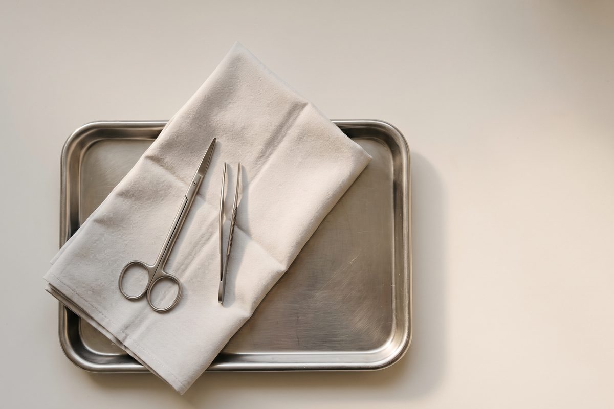
It removes it, in most cases. The methods set out in the reference sources are cryotherapy, snip excision with scissors, electrosurgery or diathermy, ligation, and radiocautery, which the reference says most specialists prefer for ease of use and precision. Lasers appear on that list twice. A carbon dioxide laser is called effective for smaller lesions and limited on large ones. A 532 nm lithium triborate laser is called a bloodless, dressing-free approach that is less effective overall than scissor excision.
That last clause deserves a moment. The comparison in the text is a laser against a pair of scissors, and the scissors come out ahead on effectiveness. Snip excision is credited there with better wound healing, less discomfort and higher patient satisfaction, with the caveat that these lesions bleed freely once cut. A clinic that owns a laser has an incentive to reach for it. The reference literature does not share that incentive.
A second reason concerns the method itself. Cutting a growth off leaves a specimen. Vaporising, burning or freezing it does not. If a lesion turns out to be one that warranted a pathologist, a destructive method has left nothing to send. How often that matters is not a number I have, and inventing one would be worse than the gap.
The four things that should send you to a doctor
The NHS version runs to four items. See a doctor if one gets bigger, becomes painful, starts bleeding, or if a lot of them develop. None of those means something is wrong. Each means the looking should be done by someone qualified.
The fourth carries information that has nothing to do with skin. Clinical references correlate these growths with type 2 diabetes and insulin resistance, obesity and central weight, abnormal lipids, polycystic ovary syndrome in younger women, and markers of cardiovascular risk. Correlation is not causation: a crop of new tags diagnoses nobody with anything, and removing them changes no metabolic number. It is a prompt to mention the finding to your own doctor and have ordinary blood work done.
What removal can go wrong with
Every method on that list has a complication profile. The clinical reference lists scarring where removal is done improperly, irritation at the site, rare bleeding, rare infection, and a neuroma with ongoing pain where a nerve running inside the growth has been cut through. Freezing adds a patch of altered colour where the cold does not stop neatly at the lesion. The same text advises a medication history first, because several drugs change how a small wound bleeds and clots.
The NHS states this plainly. Do not remove one yourself unless a doctor has told you to. The risks are infection, bleeding and scarring, with nothing on the other side. The NHS does not fund removal, classing it as cosmetic surgery, which says a good deal about how benign the typical one is, and nothing about whether yours was examined.
The one on the eyelid is a different question
These grow on eyelids, and an ophthalmology teaching resource written by oculoplastic surgeons notes that the eyelid ones are what patients notice. Diagnosis there is clinical too, and histology is rarely obtained. Lesions that mimic one on a lid include squamous papillomas, viral warts, benign melanocytic naevi, neurofibromas and seborrhoeic keratoses. One more matters: squamous cell carcinoma is rare in that position, and it is sometimes found at the base of what looked like an acrochordon. At the base of it. That matters to anyone about to burn a growth off a lid, and the same resource warns against home remedies there.
This clinic does not diagnose or treat skin cancer, does not biopsy, and does not operate on eyelids. Anything on a lid goes to an oculoplastic surgeon, an ophthalmologist with further training in the eyelids, the tear system and the socket. Dr Catherine Chow, a consultant oculoplastic surgeon and a colleague I have published with on periocular injection, has written on the eyelid skin changes that are not normal ageing, and when one should be looked at. What that assessment involves is hers to describe. The rule on this side sits in why a lesion on the eyelid is never treated as a pigment target, alongside the findings that end an aesthetic consultation here.
The question people ask second
What arrives in the room is almost always a question about method. Laser or cautery, will it scar, can it be done today. Those are the second questions. The first is whether anyone qualified has looked closely enough to say the thing is not something else. No device answers that one.
Sometimes the answer is that nothing needs doing, since some patients do not need a treatment. A harmless growth that does not catch on a collar is allowed to stay where it is. What it may not do is come off, by any instrument, before somebody has decided what it was.